Your anatomy: Urinary bladder
Provided by
Our bodies are amazing, and incredibly complex, so when things like urinary tract infections, yeast infections or occasional bladder control issues happen, it can be a little confusing as to just what is going on — and, let's be honest, they can be a little troubling when we're in the dark about how our body works. Knowledge is power, ladies, so prepare to get a little more powerful. Allow us to introduce you to: your bladder!
The anatomy of a healthy bladder
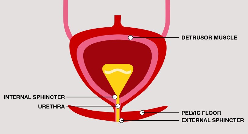
The healthy bladder is a way station, a holding area for urine (urea, water and other waste materials from the kidneys). It's a pliable, muscular organ that expands and contracts. The muscle in the wall of the bladder is the detrusor muscle. It relaxes to allow urine collected from the kidneys to fill the bladder, and then it contracts when it's time to empty the bladder.1 The detrusor is stimulated by sacral nerves,2 which are part of the parasympathetic nervous system.3 That means the detrusor doesn't rely on the participation of your conscious mind — “auto-pilot” is at work here.
Which is a good way to think about the lower part of your urinary tract (specifically the bladder and urethra) — some parts work only when you tell them to, while other parts do their work automatically. Remember, the detrusor is controlled by auto-pilot. It relaxes to allow more urine to collect, and then contracts to expel that urine through the urethra — all without you consciously controlling it.
The bladder and the pelvic floor muscles
Although the detrusor is a muscle that's actually part of the bladder, there are a host of other muscles that aren't part of the bladder itself but which are critical in helping the bladder do its job properly. The bladder is supported by the pelvic floor, an intricate network of muscles that acts like a hammock for the urinary bladder and other internal organs.4 Through the pelvic floor, the urethra passes from the bladder to the outside of the body. This tube allows urine to be expelled, but it also has a couple of muscles to help control when urine is expelled and at what rate.
The internal sphincter is part of the pelvic floor. It's also controlled by the parasympathetic nervous system (so it's also on auto-pilot). It's at the top of the urethra. Think of the internal sphincter as the throttle, controlling the rate at which urine is expelled. Below it is the external sphincter, also part of the pelvic floor. This muscle serves as the tap, and it's under your control.1 You decide when to contract it (when you're looking for the bathroom) and when to relax it (when you've finally found the toilet).
This is another way to understand your bladder muscles — some (the detrusor) are typically in a relaxed state, except when expelling urine. Others (the internal and external sphincters) are typically in a contracted state, except when expelling urine.
Support bladder health
This is how a healthy bladder functions. But like any part of the body, the bladder ages over time and can be affected by other physical changes throughout life. Fortunately, you have options. Bladder muscles are consciously controlled with bladder control exercises (Kegels, anyone?). And bladder muscle tissue that runs on autopilot can be supported with bladder health supplements. Knowing which muscles are where, and how to take care of them, is an important step toward maintaining good bladder health.
Like we said — knowledge is power. At AZO, we want to give you the knowledge you need for the power you deserve.